P.E.T.S. Systems (Percutaneous EpiphysiodesisTransphyseal Screw)
Dr. Atul Bhaskar, Dr. Avi Shah, Dr. Khyati Gupta
7/20/20244 min read
Indications:
Cancellous screws (UNIVERSAL ORTHOSYSTEMS) are designed for use in cancellous and trabecular bone, commonly found at the ends of long bones such as femur and tibia. The screws are full threaded and are available in various diameters (4 mm, 6.5 mm and 7.3 mm) with varying length (35 mm to 100 mm). It has various indications such as:
• Femoral neck fractures
• Angular deformities around knee
• Slipped capital femoral epiphyses
• Tibial plateau fractures
• Ankle arthrodesis
• Pediatric femoral neck fractures
• Intercondylar femur fractures
• Sacroiliac joint disruptions
• Subtalar arthrodesis
The key features of the screws include
• Deep cutting threads with a large pitch to increase resistance to pullout.
• Self-drilling, self-tapping screw tip facilitates screw insertion by eliminating the need for predrilling and tapping in most cases.
• Low-profile head helps reduce possibility of soft tissue irritation.
• Reverse-cutting flutes assist in screw removal.
We will be describing one of the indications of cancellous screws which is hemi epiphysiodesis for knock knees (genu valgum)
KNOCK KNEES (GENU VALGUM):
Angular deformities of the knee at the coronal plane are relatively common causes for pediatricorthopedic surgeon referrals. They cause cosmetic problems, gait disturbances, joint instabilities with pathologic crepitus, and progressive activity-related pains in growing children. Malalignment of the knee produces pathologic stress on the uni-compartmental joint space of the knee, increasing the risk of accelerated degenerative changes. The current recommendation addresses lower limb angular deformities using mechanical axis assessment as early as possible to prevent consequences such as altered gait patterns, early articular cartilage degeneration, and soft tissue changes.
Asymmetrical suppression of the physis in growing children has been proven to correct coronal angular deformities of the knee. Percutaneous epiphysiodesis using transphyseal screws (PETS) can be a good treatment option for correcting coronal angular deformities because it gradually corrects deformities and can be terminated by screw removal after completion. Measuring the average rate of correction is important when predicting the time for screw removal, and it allows patients and surgeons to expect when treatment will end.
SURGICAL TECHNIQUE FOR PET SCREWS:
1. The patient is placed in a supine position on the operating table after being given general anesthesia.
2. A radiolucent operating room table and a fluoroscope are used without the placement of a tourniquet.
3. The knee is flexed 20o and a guide wire is drilled from caudal to cranial direction from medial to lateral, crossing the medial physis from the central 1/3 in AP and lateral views of the fluoroscope.
4. The guide wire is drilled out from the lateral thigh upto the medial edge of the femoral condyle.
5. A small 1 cm incision is made and the size of the screw is measured using the device.
6. The proximal cortex of the bone is drilled using appropriate size drill broach through the sleeve to prevent soft tissue damage, up to the physis to make a tunnel for screw insertion.
7. Appropriate size (4 mm, 6.5 mm or 7.3 mm) full threaded screw (UNIVERSAL ORTHOSYSTEMS) is inserted under fluoroscope guidance, till atleast 4 threads of the screw cross the physis to achieve transphyseal compression.
8. Closure of the skin is done and a dry sterile dressing is applied.
9. Child is allowed walking on the same day/next day and to avoid any contact sports/jumping for 4 weeks post-surgery.
10. Regular follow-up is done to monitor correction of the deformity.
Screws function by converting the tightening torque into internal tension in the screw and elastic reactions in the surrounding bone. This creates compression at the physis. Screw is typically inserted into holes drilled equal to the root diameter and are either self-tapping or are inserted tapped (threaded) holes.
The torque to insert cortical bone screws can be high, so the screws must be properly inserted into the correct size drilled hole and designed to withstand insertion torque levels expected in cortical bone. Because of the relatively low strength of the cancellous bone, failure of the screw itself during insertion is rare, but pull out can sometimes be difficult.
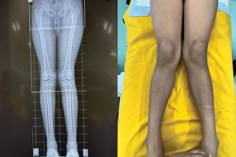
CASE EXAMPLE:
A 14 year male child, presented with bilateral genu valgum with unfused physis of the distal femur. He underwent bilateral distal femur medial epiphysiodesis with 6.5 mm full threaded Cancellous screw (UNIVERSAL ORTHOSYSTEMS).
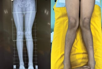
Regular follow up was done and the deformity corrected over 10 months and mechanical axis was well maintained. The screws were removed to prevent the knees to develop genu varum deformity.
REFERENCES:
1. Blair VP, Walker SJ, Sheridan JJ, et al. Epiphysiodesis: a problem of timing. J PediatrOrthop. 1982;2:281–284. 10.1097/01241398- 198208000-00007. [Abstract] [CrossRef] [Google Scholar]
2. Mesa PA, Yamhure FH. Percutaneous hemi-epiphysiodesis using transphyseal cannulated screws for genu valgum in adolescents. Journal of Children's Orthopaedics. 2009 Oct;3(5):397-403. DOI: 10.1007/ s11832-009-0203-8. PMID: 19756807; PMCID: PMC2758183
3. Abdelaziz, Ahmed MD, PhD*; ElAshry, Sameeh M. MD*; Awadh, Mohammad M. MD*; Khaja, Aliaa MBChB (Hons), KBOS†; Alsaifi, Saleh MBBCh, SB-ORTH‡. Efficacy of Percutaneous Retrograde Transphyseal Guided Growth Screw in Distal Femoral Angular Deformity Correction: A New Technique. Journal of Pediatric Orthopaedics 41(7):p e533-e539, August 2021. | DOI: 10.1097/ BPO.0000000000001835
4. Dodwell ER, Garner MR, Bixby E, Luderowski EM, Green DW, Blanco JS, Widmann RF. Percutaneous Epiphysiodesis Using Transphyseal Screws: a Case Series Demonstrating High Efficacy. HSS J. 2017 Oct;13(3):255-262. doi: 10.1007/s11420-017-9549-5. Epub 2017 Apr 3. PMID: 28983218; PMCID: PMC5617815.
5. Lee, S.-W.; Lee, K.-J.; Cho, C.-H.; Ye, H.-U.; Yon, C.-J.; Choi, H.-U.; Kim, Y.-H.; Song, K.-S. Affecting Factors and Correction Ratio in Genu Valgum or Varum Treated with Percutaneous Epiphysiodesis Using Transphyseal Screws. J. Clin. Med. 2020, 9, 4093. https://doi.org/ 10.3390/jcm9124093